Login
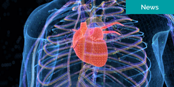
Cardiovascular Imaging
Nuclear Cardiology
Nuclear medicine is used in the diagnosis and management of diseases.
What is a Nuclear Cardiology?
What to Expect During a Loop Monitor Test
Nuclear medicine uses a very small amount of radioactive materials-called radiopharmaceuticals-to diagnose and treat disease. Radiopharmaceuticals are substances that are attached to specific organs, bones or tissues.
The radiopharmaceuticals emit gamma rays that can be detected externally by special types of cameras: gamma or PET cameras. These cameras work in conjunction with computers to form images that provide data and information about the area of the body being imaged.
MUGA Scan
A MUGA scan, which stands for “multiple gated acquisition,” is a nuclear-medicine test that allows your doctor to see if your heart is beating properly.
In a MUGA scan, a radioactive substance called Technetium 99 is is injected into the bloodstream. This substance attaches to red blood cells, and it can be viewed through the body using a special camera that ultimately produces a moving image of the beating heart.
A MUGA scan allows your doctor to measure how much blood is pumped by the heart with each heart beat. This measurement, called the left ventricular ejection fraction (LVEF) is a good measurement of overall heart function.
To schedule an appointment, contact the location most convenient to you.
Myocardial Perfusion Test
A myocardial perfusion test is a nuclear-medicine study used to evaluate the adequacy of blood supply to the heart muscle. It is similar to a routine cardiac stress test, but it also includes the injection of a radioactive tracer into the bloodstream, which enables the doctor to see more detailed information about blood flow within the heart.
This test is also sometimes called a Cardiolite stress test or a thallium stress test. Cardiolite (sestamibi) and thallium are the two radioactive substances most commonly used in this test. These are radioactive tracer materials, not a dyes. Short-term safety studies have been performed and show a large margin of safety for these substances.
Preparing for a myocardial perfusion test
If you are scheduled for a myocardial perfusion test:
- The test will take 2 to 2.5 hours.
- Wear comfortable clothes and walking shoes.
- Do not eat or drink anything with caffeine for 24 hours prior to test. This includes coffee, cola, teas, chocolate milk, chocolate pudding and chocolate candy. This also includes decaffeinated coffee, cola and tea, because these beverages still contain some caffeine.
- Do not eat or drink anything for at least 4 hours before the test. If you are diabetic, you may have juice and toast, cereal or graham crackers 2 hours prior.
- Remain on all medications unless otherwise directed by your physician. Bring a list of these medications with you.
- This test may not be appropriate if you are pregnant, suspect you may be, or are a nursing mother. Please discuss with your doctor before having this test.
- Request that a copy of your most recent EKG and prescription be sent or faxed to your appointment location by your doctor.
- We will need to know your weight before starting this procedure.
- An IV will be started for this procedure.
- If your test will include Dobutamine, please also refer to Preparing for a Dobutamine stress echocardiogram.
What to expect during a myocardial perfusion test
You will be asked to sign a consent form for the exercise part of the test. Please read the form carefully. If you have any questions, do not hesitate to ask. A physician or supervising nurse or technician will explain the entire procedure before beginning the Myocardial Perfusion Test.
Several adhesive patches, called electrodes, will be placed on your chest. These will be connected to an electrocardiographic (ECG) monitor so that your heart rate and rhythm can be watched closely throughout the test. An intravenous line will be inserted in your arm. This will be used to inject the radioisotope tracer at maximum exercise. The IV will be removed after completion of the test.
You will exercise by walking on a treadmill. This treadmill will be started at a very slow speed, and as the myocardial perfusion test proceeds, the speed and the incline of the treadmill will be gradually increased. As exercise increases, your heart rate and blood pressure will rise, which is normal. Your heart rate, blood pressure and ECG will be monitored throughout the test.
If you are unable to exercise on a treadmill, you may be given a drug called Persantine or Dobutamine, which will increase your heart rate without exercise.
You will be carefully monitored during the test. To increase the effectiveness of the myocardial perfusion test, it is important to exercise as much as you can. If you experience any unusual symptoms, such as chest pain or arm pain, shortness of breath or lightheadedness, you should tell the physician, supervising nurse or technician right away. Depending on your symptoms, blood pressure, EKG or the degree of fatigue you develop, adjustment will be made to the exercise portion of the test.
One minute prior to the end of exercise, the radioisotope tracer will be injected through the IV line. As the tracer enters the bloodstream, it is carried directly to the heart. The tracer will be visualized by a special camera that can detect radiation.
Your ECG, heart rate and blood pressure will be monitored for a few minutes after the exercise portion of the test is completed.
The technologist will position you under the camera and begin taking pictures. You will be lying on your back with your left arm over your head. The camera will be moved very close to your chest. You may receive either a Planer or a SPECT procedure. The Planer technique involves three images at different angles. Each picture will take about five minutes. During a SPECT procedure, the camera moves slowly around your chest in an arc-like fashion. The camera will acquire an image for 30 to 45 seconds and then it will move. The entire SPECT procedure will last approximately 20 minutes. It is very important to lie completely still while the pictures are being taken.
After the first set of images has been completed, you will undergo a stress test and then return for a second set of images. This will allow the doctor to compare your heart under stress and at rest. The technologist will inform you about restrictions between the two sets of pictures.
This is usually the end of the test. Our physician will discuss the results of the test with your personal physician and submit a written report. Your personal physician will then explain the results of the test and their implications to you.
Contact Us
To schedule an appointment, contact the location most convenient to you.
Related Content