

Login
Insights & Innovations - Neurosciences
The latest advancements and breakthroughs in Neurosciences
Spring 2026 Issue
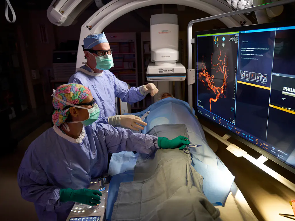
When a patient suffers a devastating brain injury or neurologic crisis, specialized expertise can mean the difference between recovery and lasting disability. At ChristianaCare’s Neurocritical Care Unit (NCCU), the only dedicated neuro-intensive care unit in Delaware, patients receive around-the-clock, cutting-edge care delivered by a highly trained, multidisciplinary team.
The NCCU features an 18-bed ICU supported by a six-bed transitional neurology step-down unit. Together, these facilities provide continuous care for patients with the most complex neurologic and neurosurgical conditions. The unit is staffed 24/7 by attending physicians, advanced practice clinicians (APCs), nurses, and therapists, ensuring seamless coverage and continuity of care.
On any given day, the team cares for patients recovering from acute ischemic stroke treatments such as thrombolysis and mechanical thrombectomy, as well as those with malignant cerebral edema, subarachnoid and intracerebral hemorrhages, and traumatic brain and spinal cord injuries. They also manage patients with neuromuscular crises related to conditions such as Guillain-Barré syndrome or myasthenia gravis who require ventilatory support.
“We are busy all the time—routinely at full capacity and sometimes overflowing into other units,” said Jennifer Creed, M.D., Ph.D., the medical director of the NCCU. “These are the sickest neurological patients in the state, and we provide them with focused, specialized care that can’t be delivered anywhere else in Delaware.”
ChristianaCare first developed the NCCU more than a decade ago, transitioning from a mixed critical care environment to a fully dedicated, closed unit as demand for specialized neurologic care grew. Dr. Creed, who stepped into leadership in 2025, now oversees the program and its expansion. Her colleagues include a team of young dynamic neurointensivists trained at premier institutions such as Thomas Jefferson, Yale, and Boston University. Together with experienced APCs and an exceptional nursing team, they’ve created a culture of teamwork and support.
“They bring fresh energy, innovative ideas, and research interests that continue to push our program forward,” Dr. Creed noted.
ChristianaCare is nationally recognized as a high-volume center for mechanical thrombectomy, ranking among the busiest programs in the country. Every patient who undergoes this lifesaving procedure comes through the NCCU for intensive monitoring and recovery.
The NCCU’s stroke program also stands out because of its aggressive approach to patients with large vessel occlusion. “Our interventionalists are willing to take on patients that many other centers would turn away,” Dr. Creed explained. “That philosophy has given many patients opportunities for recovery that wouldn’t have been possible, even a few years ago.”
Newer therapies are changing the standard of care for individuals with subarachnoid hemorrhage, a life-threatening condition often caused by ruptured aneurysms. “We’re using intrathecal medications and intravenous agents to prevent vasospasm in ways that weren’t available just a few years ago,” Dr. Creed said. “We’re practicing at the cutting edge of what’s possible for these patients.”
The unit is also contributing to the future of neurocritical care through clinical research. ChristianaCare recently began enrolling patients in the CO-MIND trial, which is developing a noninvasive device for measuring intracranial pressure (ICP). Currently, ICP requires invasive devices such as external ventricular drains or bolts.
“This device uses two scalp electrodes to simultaneously get information through mobile interactive real-time streaming technology and try to correlate that with ICP readings from the extraventricular drain and blood pressure readings from the arterial line,” Dr. Creed explained. “If this technology proves effective, there may be a role for using this noninvasive method of measuring ICP.”
In addition to enhancing neurocritical care with research, ChristianaCare leaders are exploring ways to broaden the NCCU’s reach. Plans include increasing transitional neurology capacity and extending services beyond the main campus through tele-neuro ICU consults at companion institutions such as Wilmington Hospital and Union Hospital.
“We’re always trying to think about how we can expand our services beyond the walls of the hospital to deliver neurocritical care expertise across the region,” Dr. Creed explained. “Telemedicine and consultative outreach can ensure that patients in Delaware and nearby communities get timely, expert guidance wherever they present. Patients and families can be confident that they’re in the hands of a team fully committed to delivering the highest level of neurologic care.”
When patients in Delaware and the surrounding region face the daunting diagnosis of a brain tumor, they don’t have to travel to Philadelphia or Baltimore for world-class surgical care. ChristianaCare’s Helen F. Graham Cancer Center & Research Institute offers the same advanced surgical technologies and expertise found at the nation’s top academic medical centers, delivering world-class care to the vast majority of brain tumor patients right here in Delaware.
One of the most exciting advances now available at ChristianaCare is Quicktome, a next-generation connectomics platform that brings precision and safety to brain surgery. This powerful technology enables surgeons to visualize the brain’s intricate speech, motor, visual, and cognitive pathways in relation to a tumor, creating a personalized “map” for each patient.
By identifying and preserving these critical fiber tracts, neurosurgeon Pulak Ray, M.D., and his team can chart safer surgical approaches, minimize the risk of functional loss, and successfully treat tumors in regions once considered inoperable. With Quicktome, patients gain access to surgical options and outcomes that match the nation’s most advanced neurosurgical centers—without leaving Delaware.
“In the past, there were areas of the brain where we would hesitate to remove tumors because of the risk of causing major deficits,” Dr. Ray explained. “Now, with connectomics, we can plan more precise surgeries and preserve essential functions.”
For patients with tumors near speech or motor areas, ChristianaCare offers awake craniotomy. After sedation and surgical exposure of the brain, patients are gently awakened and interact with a speech therapist during the procedure to perform simple tasks such as speaking, moving a hand, or naming objects. This real-time interaction gives surgeons a live map of language and motor function in the brain, allowing them to precisely navigate around critical regions while still pursuing aggressive tumor removal. The result is greater safety, preservation of quality of life, and the possibility of successful surgery in cases that might otherwise be considered too risky.
Another innovation now offered at ChristianaCare is 5-ALA (Gleolan) fluorescence-guided surgery. Patients receive a compound that causes high-grade gliomas to fluoresce under a special light, helping surgeons to distinguish tumor from healthy brain tissue in real time. During surgery, when illuminated with a particular wavelength of light, these tumor cells glow bright pink, allowing surgeons to see the boundary between tumor and healthy brain tissue in real time. This enhanced visibility helps the surgical team remove as much of the tumor as safely possible while preserving normal brain function, improving outcomes and giving patients a better chance at long-term recovery.
Another innovation now offered at ChristianaCare is 5-ALA (Gleolan) fluorescence-guided surgery. Patients receive a compound that causes high-grade gliomas to fluoresce under a special light, helping surgeons to distinguish tumor from healthy brain tissue in real time. During surgery, when illuminated with a particular wavelength of light, these tumor cells glow bright pink, allowing surgeons to see the boundary between tumor and healthy brain tissue in real time. This enhanced visibility helps the surgical team remove as much of the tumor as safely possible while preserving normal brain function, improving outcomes and giving patients a better chance at long-term recovery.
“On MRI, tumor and normal brain can look very similar,” Dr. Ray noted. “Fluorescence gives us cellular-level clarity, helping us achieve more complete resections.”
ChristianaCare’s neurosurgical teams also use advanced intraoperative ultrasound to differentiate tumor from swelling during surgery.
In addition, all brain tumor samples undergo next-generation sequencing, which can reveal genetic markers that open the door to targeted therapies or immunotherapies that traditionally have not been used for brain tumors.
“We are able to personalize treatment in ways that weren’t possible even a few years ago,” Dr. Ray said.
ChristianaCare’s neuro-oncology program brings together neurosurgeons, neuro-oncologists, radiation oncologists, pathologists, speech and physical therapists, and oncology nurses in a tightly coordinated, multidisciplinary model. After all procedures, neurosurgical patients are cared for in the neurocritical care unit.
“The most important part of oncologic care is collaboration,” Dr. Ray noted. “From diagnosis through surgery, radiation and chemotherapy, our teams know each other well and work seamlessly to provide comprehensive, timely treatment. At ChristianaCare, we deliver the most advanced brain tumor surgery available anywhere—and we do it right here at home.”
Neurointerventional surgeons at ChristianaCare are using a state-of-the-art approach for the management of chronic subdural hematoma (SDH). SDH is a condition caused by the formation of fragile, leaky blood vessels (neovascularization) in the membranes surrounding the hematoma over a period of weeks or even months.
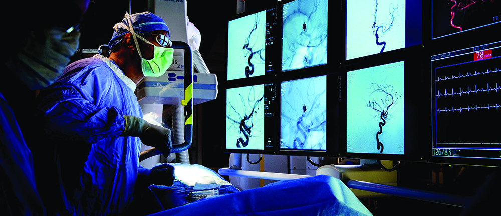
Called middle meningeal artery (MMA) embolization, the novel, minimally invasive procedure is being used to correct dysregulation of the MMA, a key artery that supplies blood to the inner surface of the skull and is prone to injury following head trauma. In many cases, the procedure is performed as an outpatient surgery, meaning patients are discharged the day of the procedure, an indication of the faster recovery times—and reduced risk for complications—associated with it.
“Historically, patients with chronic subdural hematoma have had the blood removed surgically, via craniotomy, in which a portion of the skull is removed,” said Sudhakar R. Satti, M.D., the associate director of Neurointerventional Surgery at ChristianaCare. “This is a highly invasive procedure, obviously, and the risk for complications, including recurrence of the SDH, is significant.”
Although SDH is relatively uncommon in the general population, it can have serious, even life-threatening consequences if left untreated, including seizures, stroke, coma, and death. Effective treatment strategies are therefore critically important.
Chronic SDH is most often seen in people aged 70 and older, who face a higher risk due to falls and the frequent use of blood-thinning medications to manage conditions like hypertension or atrial fibrillation. This combination of factors makes timely diagnosis and innovative treatment approaches especially vital for protecting the health and quality of life of older adults.
Developed in the early 2000s, MMA embolization is “truly a paradigm shift in how we approach chronic SDH,” Dr. Satti said.
In chronic SDH, inflammation leads to the formation of fragile, leaky blood vessels in the membranes surrounding the hematoma, and these leaky blood vessels are effectively fed by branches of the MMA, resulting in “a vicious cycle of bleeding and inflammation,” according to Dr. Satti.
The MMA embolization procedure aims to disrupt this cycle by blocking the blood supply to these abnormal membranes using a catheter that is inserted into an artery (typically via the wrist or groin) and guided to the MMA using x-ray imaging. Once the catheter is in place, an embolic agent is injected to permanently block the artery.
By targeting the abnormal vessels, MMA embolization addresses the underlying mechanism of SDH formation and growth by preventing the re-accumulation of blood and promoting the absorption of the existing hematoma by the body. Careful anatomic assessment during the procedure, again via x-ray imaging, is vital to avoid embolization of vessels that supply cranial nerves or the ophthalmic artery and causing related complications.
“Because of the high risk for recurrence associated with drainage of the SDH via craniotomy, many patients ultimately had to have multiple surgeries to resolve the issue,” Dr. Satti explained. “Research suggests MMA embolization reduces the likelihood of SDH recurrence and reduces the need for additional surgery by over 50%.”
Currently, MMA embolization is used in patients with chronic SDH and mild to moderate symptoms, either as a stand-alone treatment or as an adjunct to craniotomy (the latter being recommended for those at high risk for recurrence). However, its success in those with chronic SDH has led researchers to explore using the approach for acute and subacute—or more fast-developing—SDHs, although that is likely still a few years away, according to Dr. Satti.
“For patients with suspected chronic SDH, it’s vital that they be referred to us for treatment as soon as possible because the outcomes with MMA embolization are better earlier in the development of the bleed,” he said. “We’re seeing dozens of cases a year, many in patients who were asymptomatic or who experienced what they thought was an innocuous head trauma, and we are achieving excellent results.”
References
- J Neurointerv Surg. 2024;16(4):329-330.
- N Engl J Med. 2025;392(9):918-920.
- N Engl J Med. 2025;392(9):855-864.
- J Neurointerv Surg. 2024;17(e1):e172-e177.
Patients with essential tremor or tremor-dominant Parkinson’s disease whose symptoms do not improve with standard pharmacotherapy have access to a state-of-the-art treatment at ChristianaCare called magnetic resonance–guided focused ultrasound (MRg-FUS).
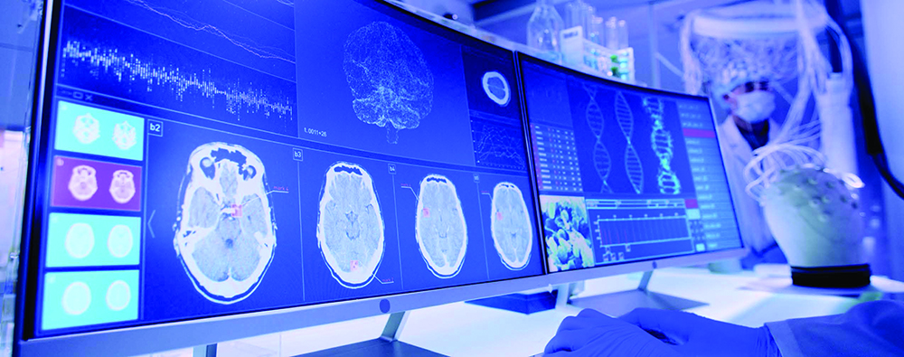
This outpatient procedure delivers high-intensity ultrasound beams through the skull to a specific target in the thalamus, without the need for anesthesia or cranial implants. Patients are awake and responsive throughout the procedure, which enables real-time assessment for efficacy as well as any potential adverse events and/or complications.
He continued: “Essential tremor and tremor-dominant Parkinson’s disease are conditions neurology has treated for many years with medications but, unfortunately, medications are only about 60% to 70% effective at reducing tremor in the best studies. Focused ultrasound provides an excellent option for patients who are refractive to medical intervention, one that is safe, effective, and accurate.”
Essential tremor is one of the most common movement disorders in older adults, affecting up to 9% of those aged 60 years and older.1 Parkinson’s disease is less common, affecting more than 1 million adults in the United States—most of them aged 60 years and older. However, tremor is a dominant symptom in up to 75% of those with the disease.2 These figures are significant, given the aging population in Delaware.
“We know there are a lot of retirees living in Delaware and, if they are experiencing tremor, it can have a significant impact on their quality of life and their ability to maintain independence,” Dr. Ray said. “For those who are refractory to medication therapy, tremor can become debilitating, affecting their ability to perform daily activities.”
Deep brain stimulation (DBS) has previously been considered the standard of care for the neurosurgical treatment of essential tremor and tremor-dominant Parkinson’s in patients who do not respond to medication. Although it is considered reversible and adjustable, the procedure is invasive, as it involves craniotomy and the permanent implantation of electrodes into the brain. These electrodes stimulate the target area, inhibiting neural activity and alleviating the tremor; however, they are battery operated and the batteries require periodic replacement, according to Dr. Ray.
Historically, neurosurgeons have also treated these tremor disorders with thalamotomy. Because of the invasive nature of the procedure, however, surgical thalamotomy may be inappropriate for many older adults, many of whom are frail and have underlying cardiovascular disease and other health problems, according to Dr. Ray.
Both DBS and surgical thalamotomy carry significant risks, typically adverse effects related to inaccurate target localization, including ataxia, paresthesias, dysarthria, hemorrhage, and/or seizures.
MRg-FUS is essentially a noninvasive form of thalamotomy. MRI provides detailed images of the brain in real time during the procedure, enabling precision in identifying the target area treatment, which differs from patient to patient, and minimizing risk to the surrounding tissue. High-intensity FUS is delivered to a focal point through the intact skull using a framing device that is placed on the patient’s head. Patients remain fully conscious during the procedure—no general anesthesia is required—and its functional effects are assessed throughout, allowing the multidisciplinary clinical team to refine and confirm the correct target and appropriate number of sonications.
Typically, tremor improvement is immediate, and the noninvasive nature of the procedure yields faster recovery and lower risk for complications such as hemorrhage and infection, Dr. Ray said.
“Focused ultrasound gives us the accuracy needed to stun the cells in the areas of the brain that cause the tremor,” he noted. “As a result, the efficacy is remarkable, with up to 73% of patients still seeing a reduction in tremor at five years.”
The entire procedure takes less than two hours—from patient arrival to discharge, according to Dr. Ray. It is performed in the MRI suite, as opposed to a traditional operating room. All patients are cared for by a multidisciplinary team that ensures safety and comfort before, during, and after the procedure.
Although medication is still first-line therapy for both essential tremor and tremor-dominant Parkinson’s disease, MRg-FUS has become a viable option for patients whose symptoms do not respond to treatment, even those who have other underlying health conditions. Patients with bilateral tremor can have both sides treated via the approach, in procedures scheduled nine months apart.
“Many of the patients who have these tremor disorders are quite frail because they’re elderly, so the ability to provide noninvasive neurological procedure is significant, and we have achieved excellent results,” Dr. Ray said. “At ChristianaCare, our ability to provide this less invasive, innovative treatment for this debilitating condition is really a unique benefit that will serve patients in our community and throughout the region.” •
References
- Zappia M, Albanese A, Bruno E, et al. J Neurol. 2012;260:714-740.
- Pasquini J, Ceravolo R, Qamhawi Z, et al. Brain. 2018;141(3):811-821.
For patients living with chronic neurologic conditions such as epilepsy and chronic headache, regular follow-up visits are an essential part of care. Yet mobility challenges, driving restrictions and the unpredictability of symptoms often make it difficult to attend in-person appointments. At ChristianaCare, neurologists are embracing virtual care to improve access, reduce barriers, and deliver high-quality treatment to patients wherever they are located.
ChristianaCare’s headache team began offering video visits at the onset of the COVID-19 pandemic. What started as a necessity quickly revealed lasting benefits. “Patients love the convenience,” explained neurologist and headache specialist Jessica Bradley, M.D. “Some of them may live up to two hours away; and for those with neurologic conditions that make driving difficult, a virtual visit saves the challenge of arranging transportation. Others can more easily fit an appointment into their workday or between college classes. It just makes care more accessible.”
For migraine patients, telehealth has been especially valuable. “If you’re in the middle of a migraine, driving and sitting in a bright office can be unbearable,” Dr. Bradley noted. “With video visits, patients can stay home, turn the lights down and still get the care they need.”
Virtual visits also have reduced missed appointments because patients can log in even when they’re not feeling well enough to travel.

Dr. Bradley emphasized that new or concerning symptoms, such as new visual disturbances or vertigo, still require an in-person evaluation, but she encouraged patients to take advantage of video visits for routine follow-up and medication adjustments when appropriate.
“For headache care, even new patients can benefit from a virtual first visit, since diagnosis is so heavily based on history,” she said.
For people with epilepsy, virtual care can be even more transformative. Jonathan Pollard, M.D., the director of the ChristianaCare Epilepsy Center, listed the multiple daily obstacles many of his patients face: inability to drive due to seizures, financial strain from job loss, and reliance on family members for transportation.
“In-person visits can be almost impossible for some patients,” Dr. Pollard said. “Virtual visits have been a godsend. They remove the transportation barrier and give patients a reliable way to stay connected with care.”
The American Epilepsy Society now recommends that virtual care be made available to epilepsy patients whenever possible, and more than half of ChristianaCare’s epilepsy patients take advantage of telehealth at least part of the time. Although certain assessments still require in-person visits, Dr. Pollard noted that “95% of the information we need can be obtained virtually. And given that many of these patients might have otherwise missed their visit entirely, virtual care is clearly an essential option.”
ChristianaCare is also advancing virtual care in epilepsy through the use of patient-reported outcome measures (a similar model is being used with headache patients), an approach inspired by a successful model in Denmark. Epilepsy affects about 3% of the general population, yet accounts for up to one-fourth of all neurology outpatient visits. To make the best use of limited clinic resources, ChristianaCare Neurology set out to identify its healthiest epilepsy patients and safely reduce the frequency of their routine visits, freeing up time for those who need it most.
Here’s how it works: At the end of an in-person or a virtual visit, relatively healthy patients are offered the option of returning in six months as usual or completing a patient-reported outcome measures survey at the six-month mark. The survey, delivered securely by text message, asks about seizure severity, medication side effects, disability and other relevant issues. If responses raise concern, the patient is scheduled for an immediate appointment. If results are reassuring, the next follow-up is extended safely to 12 months.
Early results have been encouraging. Among the first 76 patients enrolled, more than 60% responded to the survey—double the typical response rate seen in epilepsy populations nationally. Based on this success, ChristianaCare is now designing a similar survey for migraine patients, using the same paradigm.
“Virtual care allows us to see patients sooner, keep them engaged and prevent lapses in treatment,” Dr. Bradley said. “It’s not a substitute; it’s an enhancement.”
Advanced practice clinicians (APCs) play an integral role in the evaluation and management of ChristianaCare patients with neurologic disorders, with the ultimate goal of expanding access to treatment services for those with acute and chronic neurologic illness.
The Neurosciences service line’s APC program has expanded significantly over the past 15 years, growing from two APCs to 48 in that period. These specially trained clinicians assist in the provision of a wide range of services across the patient care continuum, working in four services: inpatient, outpatient, neurocritical care and neurointerventional surgery.
Although neurologists collaborate in the diagnosis and treatment for all patients, APCs serve as front-line providers, ensuring patients and their caregivers have access to all needed services and resources.
“In general, the Neurosciences service line here at ChristianaCare has a very high level of respect for APCs, which is why we’re working at the top of our license,” said Erin Mitchell, DNP, APRN, AGACNP-BC, FNP-BC, CNRN, SCRN, who is the chief APC of neurosciences at the health system. “We are truly an integral part of the award-winning care offered here.”
Neurology APCs are licensed healthcare professionals—such as nurse practitioners, like Mitchell, and physician assistants—with training in the diagnosis, treatment and management of neurologic disorders. At ChristianaCare, these professionals work collaboratively with neurologists to provide comprehensive care to patients from initial evaluation (i.e., collecting medical histories, performing physical and neurologic exams, and ordering and interpreting diagnostic tests) to disease treatment and management.
In conjunction with the health system’s award-winning neurologists, APCs develop and implement treatment plans, prescribe medications, and manage neurologic conditions, such as stroke, epilepsy, Parkinson’s disease and multiple sclerosis, according to Mitchell, who has been at ChristianaCare since 2009.
They are also actively engaged in patient and caregiver education, helping to boost understanding of complex neurologic conditions, treatment options and self-management approaches.
Because of its extensive onboarding and orientation process, ChristianaCare’s neurology APCs deliver important care, performing procedures such as lumbar punctures, nerve blocks, invasive line placement, skin biopsy for evaluation of certain neurologic conditions, and OnabotulinumtoxinA (Botox, Allergan) injections for those with migraine.. In addition, they serve as key clinicians within the health system’s stroke centers (assisting with evaluations and post-stroke care) and its epilepsy monitoring unit, where they assist in evaluating seizures and adjusting medications.
Many of the 48 APCs at ChristianaCare have subspecialties such as critical care, movement disorders, stroke, and headache, among others, according to Mitchell.
“ChristianaCare offers APCs robust training; it’s almost like a fellowship,” she said. “Our onboarding and orientation ranges from 12 weeks for our inpatient neurology APCs to up to 20 weeks for APCs working in neurocritical care. It’s very demanding, and it ultimately enhances the care we offer patients. The majority of our APCs come to us right out of school and stay, which speaks to the strength of our process.”
Fully integrated APCs offer more than just improved patient care. They improve efficiency in the delivery of patient care by freeing up neurologists to focus on more complex cases, as APCs manage routine services. APCs also enable 24/7 care by handling overnight and weekend hours in the neurocritical care unit.
“APCs help make sure that all of the T’s are crossed and the I’s are dotted,” Mitchell explained. “In general, all of the inpatient neurology patients at ChristianaCare see the attending neurologist for their initial consultation, our APCs have the training needed to essentially take over from there. Because of the expertise of our APCs, we now have providers available overnight to answer patient and family questions. And we can be a second set of eyes for neurologists evaluating very complex diagnoses and plans of care.
Indeed, that team approach only strengthens the care patients receive across the neurology service line and adds to the support families and patients receive, she added. NeuroInterventional Surgery APCs work with the physicians to facilitate elective and urgent procedures, improve transitions of care, and assess patients post-procedure. They are integral to ensuring that patients and families have all of the questions answered before and after the procedure.
“As APCs, we understand their diagnosis, we understand their medications, and we are able to provide an education component that patients and their families are able to understand,” Mitchell noted. “A real advantage of having an APC partner in your care is that we’re all very detail-oriented, knowledgeable, and hold ourselves to a high standard of care. We assume responsibility for things like care transitions, which can be high risk for many of our patients, whether it’s from inpatient to discharge or from service to service. We truly own those workflows no matter where patients are in the hospital system.”
In our outpatient services, APCs are an integral part of hospital follow-ups and longitudinal care for patients with chronic neurologic conditions. In addition to diagnosis and management, they provide access to procedures necessary for ongoing care. They are a key part of ensuring patients continue to receive the care they need outside the hospital and preventing readmissions.
“Patients at other health systems may start a new medication, get discharged, and never receive the follow-up they need,” Mitchell said. “If they experience side effects or need changes to their treatment, they can fall through the cracks. Our APCs help close those gaps, ensuring patients have the ongoing support and management that makes their care safer.”
Winter 2025 Issue
People who experience seizures die at a higher rate than those who don’t, and 70% of seizure-related deaths result from asphyxiation caused by turning face down on the pillow during sleep. John Pollard, M.D., a neurologist and director of the Epilepsy Center at ChristianaCare, has developed a novel technology that can prevent these incidents: a headband that can alert another person in the same residence that a person with epilepsy is lying in this dangerous position.
“It’s been shown that if you can warn someone, then they can flip the patient over and save their life,” he said, adding that the headband works in much the same way that a cellphone’s accelerometer can tell the user the location of their phone. “In the case of this device, the accelerometer is in your headband and if the headband detects that your face is down while you are asleep, then it will sound an alarm. It first has a quiet alarm to warn the person themselves that they are face down and, if they can’t or don’t respond, then it will sound a louder alarm for a loved one or caregiver.”

The headband was made possible through a collaboration with ChristianaCare’s Health & Technology Innovation Center. To fully realize the potential of new technologies designed to impact health and improve patient care, ChristianaCare’s Health & Technology Innovation Center brings together clinicians and information technology experts to harness emerging technologies and develop unique solutions for delivering the best possible care.
Staffed with a team of award-winning software developers, designers and user experience analysts, the center collaborates with physicians, nurses and other healthcare professionals with new solutions to create a better experience of care and better health.
Dr. Pollard entered his idea into the Health & Technology Innovation Center’s Innovation Challenge, a biennial event that invites care providers to submit new ideas, apps or technology designed to improve patient care, enhance community connection and address relevant health challenges. Although Dr. Pollard did not win the challenge, the staff at the center believed the idea was worth developing.
“The idea was interesting enough that they developed a prototype and there is a patent pending,” Dr. Pollard said.
This innovative headband highlights how neurosciences continue to be at the forefront of advancing science and integrating cutting-edge technology to enhance patient safety, prevent seizure-related deaths, and ultimately provide better care.
Autism spectrum disorder (ASD) encompasses a range of limitations in reciprocal and communication developmental milestones, resulting in significant functional challenges throughout life. Although there is no consensus regarding the brain structures and mechanisms underpinning ASD, some researchers have focused on abnormalities that affect the mirror neurons and other cortical areas involved in social reciprocity, while others have proposed a more widespread alteration in neuronal organization resulting in abnormal white matter trajectories that lead to cortical over- or under-connectivity.
Mitra Assadi, M.D., and her team at ChristianaCare have focused their research on a target they define as “the social reciprocity network”—the mirror neurons in the inferior frontal gyrus (IFG) and inferior parietal lobule (IPL) of the brain, as well as the cortical areas involved in abstract social cognition, including the medial prefrontal cortex, temporal-parietal junction and posterior cingulate gyrus.
“We hypothesize that aberrant connectivity affecting the above neuronal circuitry, what we call ‘the social reciprocity network,’ contributes significantly to the core deficits in this condition,” explained Dr. Assadi, a pediatric neurologist the medical director for neuroscience education at ChristianaCare.
The research team is launching a study using quantitative electroencephalography (QEEG)-based coherence and spectral power analysis to measure the functional connectivity and neuronal dynamics involving the social reciprocity network in patients with ASD. The research protocol offers study participants noninvasive brain modulation, via repetitive transcranial magnetic stimulation (rTMS), which may mitigate the aberrant behaviors in ASD by altering neuronal dynamics and connectivity.
“Our overarching goal is to explore the neuronal connectivity and oscillatory patterns in the social reciprocity network and to elucidate the mechanism of modulating the above variables in improving social cognition,” said Dr. Assadi, who is also a professor of neurology at Thomas Jefferson University.
“Everyone has a tremor center of their brain,” Dr. Martello explained. “For patients with tremors it is not functioning correctly.”
The study, for which enrollment began in the fall of 2024, will involve 37 ASD patients ages 13 to 18 years. Participants will undergo 18 sessions (three times per week for six weeks) of rTMS or sham stimulation of the bilateral IPL/IFG in a prospective, single-blind study design. During phase I (three weeks), half of the subjects will receive rTMS and the other half will receive sham stimulation. During phase II (three weeks), all subjects will receive active rTMS.
The type of rTMS used in the study, known as intermittent theta burst stimulation (iTBS), is unique in inducing the production of brain-derived neurotrophic factor, leading to neuronal plasticity. Participants undergoing active treatment will receive 2,400 stimulations per session, divided equally between the bilateral IPL and IFG. The outcome measures include a neuropsychological battery (at baseline, phase 1 and 2, and three weeks after completing rTMS) and QEEG analysis (on the first and last active treatment sessions).
“The QEEG enables us to measure the coherence and spectral densities and determine if there is a reproducible pattern within the social reciprocity network in ASD,” Dr. Assadi said. “To measure the behavioral effects of rTMS/iTBS, we will use the Childhood Autism Rating Scale as well as the Social Responsiveness Scale. We expect to see an improvement in the behavior scores commensurate with enhanced social cognition, enhancement of the beta and gamma powers, and the connectivity within the social reciprocity network.”
Dr. Assadi explained that “this exploratory research builds upon our proof-of-concept testing of 11 children with ASD who received 10 sessions of rTMS targeting unilateral IPL.” The QEEG analysis performed in four subjects demonstrated an enhancement of beta and gamma powers post-rTMS in three cases, she added.
“We also demonstrated a modest improvement in social awareness,” Dr. Assadi said. “The rTMS/iTBS targeting of the bilateral social reciprocity network is a novel concept in ASD. Analyzing the correlation between the behavioral outcomes and the alterations in the neuronal connectivity and dynamics may provide a deeper understanding of the neuroanatomical substrate underpinning ASD, and inform the future clinical trials leading to effective and personalized treatment approaches.”
A patient diagnosed with multiple sclerosis (ChristianaCare’s pediatric neurology program provides comprehensive, continuity-focused care delivered by an experienced team of physicians. The program emphasizes collaboration with families and pediatricians to develop personalized treatment plans tailored to each patient’s needs, addressing both simple and complex neurological conditions by combining advanced diagnostics, cutting-edge treatments, and decades of expertise.
“We offer treatments for a range of neurological disorders, including common conditions such as headaches, Tourette’s syndrome, epilepsy, and attention-deficit disorder, as well as more complex issues like developmental delays and autism,” said ChristianaCare pediatric neurologist Mitra Assadi, M.D., medical director for neuroscience education. “We are able to define pediatric epilepsy syndromes based on findings from standard EEG recordings, ambulatory EEG, or long-term video EEG monitoring.”

According to the American Academy of Neurology guidelines, genetic testing is a crucial component of further assessing patients with developmental delays, explained Dr. Assadi, a professor of neurology at Thomas Jefferson University. At ChristianaCare, DNA samples are obtained via a simple oral swab provided during the initial consultation.
Genetic testing proves highly impactful, leading to precision cellular or molecular treatments in 15% to 20% of cases. Even when such treatments are not available, genetic testing offers significant benefits by concluding the diagnostic odyssey for patients and their families, Dr. Assadi noted.
“Many of these genetic syndromes involve multiple systems,” she explained. “Therefore, having a genetic diagnosis allows us to put the pieces of puzzle together and understand the big picture.”
Genetic testing can also provide useful information for future family planning and prognostication. Discovering a specific mutation in the patient will allow us to perform DNA testing on the unborn fetus and determine if the unborn child is going to be affected.
“Some parents may decide to abort a baby expected to have a poor prognosis,” Dr. Assadi noted. “We also provide targeted genetic testing for conditions such as muscular dystrophy or familial neuropathies. After establishing the diagnosis, we will refer these patients to specialized centers to receive cell/gene-based treatments. In addition to providing a great service, at ChristianaCare, we are committed to continuity of care. We are experienced and seasoned pediatric neurologists with 30 plus years of experience.”
In the skies over Delaware, ChristianaCare’s LifeNet air medical transport team is pairing cutting-edge aviation technology with critical care medicine to save precious moments in the race to treat patients who have experienced stroke and other trauma.
Navigating the complexities of patient transfer, prepping care teams and ensuring maximum efficiency from the time a stroke is first recognized can have a significant impact on preventing long-term disability and death. The LifeNet team is revolutionizing stroke and trauma care in Delaware by leveraging advanced aviation technology and streamlined processes to significantly reduce transfer times, ultimately improving patient outcomes and enhancing the chances of recovery.
“It’s estimated that roughly 1.9 million brain cells die every minute that a stroke goes untreated,” said John Roussis, MBA, NRP, FP-C, CMTE, the program director for pre-hospital services at ChristianaCare. “So, early recognition and activation is key. We work with ChristianaCare’s multidisciplinary teams to constantly try to improve our metrics, and to use high-performance efficiencies to get beds ready, notify clinicians, prep our laboratories and decrease time to treatment for these patients.”
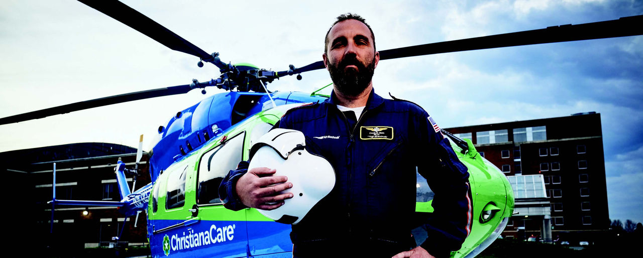
On the ground, a custom “easy button” placed in referring community hospitals allows medical staff to immediately notify the pre-hospital team anytime a case may require a time-sensitive transfer. Pressing the button engages the pre-hospital communication center, which then places a helicopter or ambulance on standby. While that happens, information regarding the case flows into the center, helping to determine the correct response, either air or ground transport, and passing along critical information to hospital emergency departments.
“Even five years ago, we would have to wait for a physician to phone the center to request a transfer,” Roussis said. “Transport—the part of the process that often takes the longest to organize—would be activated four or five tick-marks into the response process. Now, we already have transport on the way well before the physician is done assessing the patient.”
The two LifeNet helicopters—one based at Christiana Hospital in Newark and the second at Delaware Coastal Airport in Georgetown—are staffed by flight teams accredited by the Commission on Accreditation of Medical Transport Systems and available 24/7 to provide emergency air transport for critically ill or injured adult, pediatric and neonatal patients.
Pilots of the aircraft are instrument flight rules—or IFR—rated, meaning they can fly while relying solely on reference instruments, allowing them to move efficiently through areas of differing airspace and safely navigate in adverse weather conditions. Additionally, the teams are cross-trained for both ground and air, meaning they are able to avoid significant delays in the event they are unable to fly.
As a result, in Delaware, stroke patients located in the farthest reaches of the state can often be transported to ChristianaCare more quickly than was previously possible; which is critical for patients needing urgent surgical intervention for treatment of their stroke. One hospital in southern Delaware experienced an 84-minute reduction in door-in-door-out times for these transfers, and similar rates have been seen among the other referring hospitals.
These time savings can have a significant impact on patient outcomes. Studies have shown that during stroke or transient ischemic attack, the brain ages by a rate of roughly 3.6 years per hour without treatment, and the effect of stroke on cognitive function is equivalent to 7.9 years of aging.1
“Every minute you save through this process gives somebody an increased chance of having less disability and a better chance of life,” Roussis said. “If you can save three or four minutes per step in this process, that can potentially mean the difference between long-term ventilator care and being able to return to work on Monday.” •
References
- Front Aging Neurosci. 2019;11:348.
“Neuro-ophthalmology overlaps with many fields of medicine—as the name suggests, it bridges the fields of neurology and ophthalmology,” said Muhammad Asad Fraz, M.D., a board-certified outpatient neurologist and fellowship-trained neuro-ophthalmologist at ChristianaCare’s Ophthalmology Wilmington Health Center. “It focuses on all ophthalmological disorders related to the central or peripheral nervous system.”
Patients referred for neuro-ophthalmology evaluations are often medically complex and have already seen multiple specialists. Typically, patients seen by Dr. Fraz are referred either from neurology, ophthalmology, neurosurgery or otorhinolaryngology (ENT).
By the time they meet with Dr. Fraz, they are seeking expert guidance for difficult-to-diagnose conditions. This includes patients with different issues including double vision, pseudotumor cerebri syndrome (also known as idiopathic intracranial hypertension), headaches, transient visual disturbances, brain tumors, stroke, optic nerve pathology, eye movement disorders and eyelid droop.
“Patients come to me with high expectations for the best possible care, and rightly so,” he explained. “Sometimes patients with brain or orbital tumors affecting vision benefit from neuro-ophthalmology assessments that may guide surgical decisions.”
Before establishing the adult neuro-ophthalmology program at ChristianaCare, patients in Delaware often had to travel to Pennsylvania or Maryland, facing logistical and insurance challenges. Now, ChristianaCare provides these specialized services locally, eliminating the need for out-of-state travel.
“Now, we can serve the community and reduce barriers to care. It’s rewarding to know I’m making a difference,” said Dr. Fraz.
The new program actively collaborates with subspecialties across neuroscience and ophthalmology, including headache, neuroimmunology, neuro-interventional surgery, neurosurgery, retina, glaucoma and oculoplastics. Temple University ophthalmology residents also rotate in the Neuro-Ophthalmology Clinic as a part of their training.
“Working closely with colleagues in both neurology and ophthalmology enhances the care we provide,” Dr. Fraz noted.
Beyond ophthalmology, Dr. Fraz actively collaborates with other subspecialists within the ChristianaCare system. A notable example is his partnership with the neurointerventional surgery team and neurosurgery to address idiopathic intracranial hypertension (IIH), a condition that predominantly affects young to middle-aged individuals, primarily women. Patients with IIH often experience symptoms like headache, vision loss, pulsatile tinnitus and double vision due to increased intracranial pressure. This collaboration includes venous sinus stenting with neurointerventional surgery and ventriculoperitoneal shunting with neurosurgery, streamlining the patient’s treatment journey within ChristianaCare.
“In cases where medical treatment is insufficient, surgical interventions may be necessary to preserve vision,” Dr. Fraz explained. “We’re proud to be growing and bringing something new to the state. All great things have to start somewhere.”
The novel magnetic resonance (MR)-guided focused ultrasound technology allows for tremors to be controlled without surgical incisions, meaning that patients can experience immediate results with minimal to no recovery time. At ChristianaCare, the first MR-guided focused ultrasound procedure was performed on June 6, 2024. Since that time, the team has completed nearly 20 procedures.
“We’ve ramped up a lot more quickly than expected,” said Justin Martello, M.D., the director of the Parkinson’s and Movement Disorders Programs and Focused Ultrasound Program at ChristianaCare Neurology Specialists.
Although the initial plan was for the team to conduct approximately four MR-guided focused ultrasound procedures per month, the success of, and demand for, the procedure meant goals were quickly revised.
“After the first month and a half, when we saw how successful the program was and how dramatically it was improving these patients’ lives, we increased the frequency of the procedures to two per week,” Dr. Martello said.
In addition, exciting developments regarding focused ultrasound are being tested in clinical trials, likely meaning that the procedure will be able to be used in new and innovative ways in the near future. The first, which Dr. Martello noted will likely occur by the end of the first quarter of 2025, is a new indication to use the technology to treat other symptoms of Parkinson’s disease, such as slowness and stiffness of movement and dyskinesia. By mid-2025, another new indication may expand the use of focused ultrasound even further; specifically in the area of cancer diagnostics.
“This current treatment is technically considered high-intensity focused ultrasound,” Dr. Martello clarified. “And an indication for low-intensity or low-frequency ultrasound is likely.”
Whereas high-intensity ultrasound uses heat from wavelengths to target and kill specific areas of the brain that are responsible for causing tremors, low-intensity ultrasound does not heat up the brain tissue. Instead, the technique safely breaks open the blood–brain barrier for a brief period of time—often 1.5 to two hours—allowing unprecedented access to regions of the brain.
“When a patient has a tumor in their brain, often it isn’t possible to do surgery in order to either conduct a biopsy or remove the tumor due to its deep position in the brain,” Dr. Martello said. “This technique, which is called liquid biopsy, opens up the blood–brain barrier and stimulates blood flow around the tumor.”
This can slightly break up the tumor, allowing its cells to circulate in the blood. Doing so means doctors can take a blood sample, which will contain cancer cells that can then be tested in order to complete a diagnosis without necessitating surgery or traditional biopsy. While breaking up pieces of the tumor can aid in diagnosis, it does not result in spreading of the cancer to other areas of the body.
“What it does do is allow for a noninvasive way to diagnose the cancer that doesn’t risk permanent damage to the brain,” Dr. Martello said.
The technique may also allow for novel ways of delivering medications that aren’t normally able to cross the blood–brain barrier, potentially offering benefits for patients undergoing chemotherapy, as well as those receiving treatment for conditions such as Alzheimer’s disease.
These developments, Dr. Martello says, will help to both increase the use of this cutting-edge technology and allow for more neurologists to offer the benefits of focused ultrasound to their patients.
The goal of a scheduled EMU admission for a patient is to characterize and localize the seizure-like event so that the appropriate diagnosis can be made and treatment can be initiated. Many patients treated in the EMU are there for preoperative evaluation before surgery designed to control seizures, if pharmacotherapy has failed.
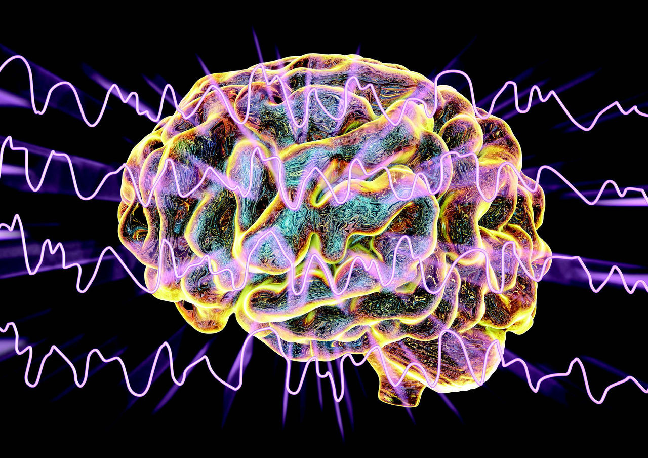
The EMU is a specially equipped inpatient unit that uses continuous EEG monitoring, continuous video monitoring through ChristianaCare Virtual Health and continuous heart rate monitoring, according to John Pollard, M.D., an adult epileptologist and the director of the Epilepsy Center at ChristianaCare. The EMU strives to provide a superior level of for complex epilepsy patients and accredited by the National Association of Epilepsy centers as a Level 3 center. Level 3 centers provide a range of neurodiagnostic evaluations and medical, neuropsychological, and psychosocial services needed to diagnose and treat patients with refractory epilepsy. Between 50 and 100 patients visit the unit annually.
“When someone has a seizure in our EMU, the nurse can come in and care for the patient and perform an assessment called an ictal cognitive exam,” Dr. Pollard said. “This combination of factors—the video, the ictal cognitive exam and the EEG exam—is very effective at determining what kind of seizures people are having.”
“In the EMU, we do presurgical evaluation; we do differential diagnosis to capture a seizure episode and make the right diagnosis, and we also use the EMU to adjust medications occasionally,” noted June Wang, M.D., PhD, an adult epileptologist and EEG lab director at the Christiana Hospital.
At least half of the people who go into the EMU at ChristianaCare don’t know why they are having seizures, and some of them turn out to be having events that are not actually epilepsy but the result of some other cause, such as a psychological problem or a heart arrhythmia that leads syncope. In some cases, a patient’s seizures are determined to be from a transient ischemic attack.
“We want to see if someone is eligible for an epilepsy surgery by trying to discover whether it would be safe to take out the spot or spots in their brain that is causing the seizures,” Dr. Pollard noted.
The EMU has also instituted what its specialists call a fall prevention system to limit the risk for the 1 in 10 epilepsy patients (national average) who experience a fall in the hospital.
“Our fall prevention system has been adapted from the system used by physical medicine and rehabilitation,” Dr. Pollard said. “It’s a vest that attaches to the bed lift, so if someone has a fall, then they don’t actually hit the ground because the vest stabilizes them. Uniquely, in our EMU, falls are incredibly rare.”
Because of these and other innovations, ChristianaCare’s Epilepsy Center of Excellence is prepared to care for a range of patients with epilepsy and seizure-like events, including those with special needs. In addition, the health system’s pharmacy has a protocol for pregnant women who often need their epilepsy medications adjusted; pregnancy alters blood levels of certain medications, placing women at increased risk for seizures.
To expedite appropriate care, the epilepsy team has developed and instituted an uncomplicated seizure pathway that provides quick treatment access to patients who present to the emergency room with seizures, and newly diagnosed patients with epilepsy are offered education and expedited ambulatory appointments. The ChristianaCare Epilepsy Center of Excellence also works closely with neuropsychology and neuroradiology to ensure accurate diagnoses, and with neurosurgeons to provide on neuromodulation therapy, if applicable.
Spring 2024 Issue
To address this issue in New Castle County, home to 60% of Delaware’s population, ChristianaCare has partnered with the county’s emergency medical services (EMS) to become an early adopter of Twiage, a novel prehospital communication and intrahospital care coordination technology. The system is designed to facilitate communication between hospital personnel and EMS to accelerate lifesaving emergency care by reducing dependence on radio communication.
Twiage is available to EMS personnel for free, and the HIPAA-compliant app can send notifications with patient vital signs, symptoms (including EKG results) and interventions to the hospital in seconds, along with photos, additional details in text chats and digital voice memos. That information is readily available to health-system vascular neurologists on their cellphones, according to Kim Gannon, M.D., Ph.D., the medical director of ChristianaCare’s Comprehensive Stroke Program and physician executive of the Neurosciences Service Line.
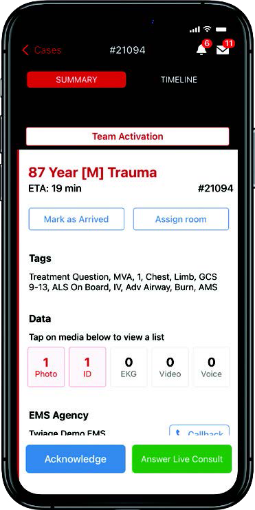
“When neurologists can have that information available, prior to the patient’s arrival at the hospital, it can make all the difference in the world because seconds truly matter in stroke care,” said Dr. Gannon. ChristianaCare’s stroke program is one of the largest nationally and one of only 300 comprehensive centers across the country. “With this system, paramedics can communicate with our stroke teams while they’re on their way to the hospital, which gives our staff time to access and review the patient’s chart. This is particularly important if the patient is unable to speak for themselves or doesn’t have a family member present.”
Research suggests that stroke patients lose approximately 1.9 million brain cells per minute during care delays, making rapid initiation of antithrombotic therapy crucial, added Doug Huisenga, the senior program manager for ChristianaCare’s systemwide stroke program. However, although these drugs are widely used and effective, there are contraindications in many patients, including a history of cerebral hemorrhage and/or ischemic stroke (within the past three months).
“That’s why having access to a patient’s personal health information is so important,” he said.
Efficient stroke care represents a particularly acute need in Delaware, which is actually located in the northernmost reaches of the so-called “stroke belt,” the region comprising most of the Southeastern United States that has the highest incidence of stroke mortality in the country. The state actually has the second-highest incidence of stroke mortality (56.8/100,000 people in the general population) in the country, behind Mississippi.
Of note, hypertension and hyperlipidemia as well as tobacco use are very common in Delaware. ChristianaCare is the only comprehensive stroke center on the I-95 corridor between Baltimore and Philadelphia.
The initiative with New Castle County EMS and ChristianaCare means Delaware is now one of 12 states nationally in which the Twiage system is in use. By providing patient data and GPS-tracked ETA for all incoming ambulances in real time, Twiage helps reduce hospital efficiency and enables earlier initiation of treatment, meaning lives are saved.
“ChristianaCare is already likely in the top 1% of hospitals nationally in door to needle time,” said Huisenga, referring to the initiation of antithrombotic treatment. “However, one of the ways we can make that even better is to improve prehospital communications — and that’s what we’ve done with Twiage thanks to this collaboration with New Castle County EMS.”
Cutting-edge magnetic resonance (MR)-guided focused ultrasound technology is allowing neurologists at ChristianaCare to provide outpatient procedures for patients struggling with essential tremor (ET) and Parkinson’s disease.
Developed in the early 2000s, and approved by the FDA in 2016, the technique allows for tremor control without surgical incisions, meaning that patients can recover quickly, are not at risk of destabilizing infections and are able to experience immediate reduction of their tremors. This summer, Justin Martello, M.D., the Parkinson’s and movement disorders director at ChristianaCare Neurology Specialists, will use the technology for ET and Parkinson’s patients as the first in the state of Delaware to offer this treatment.
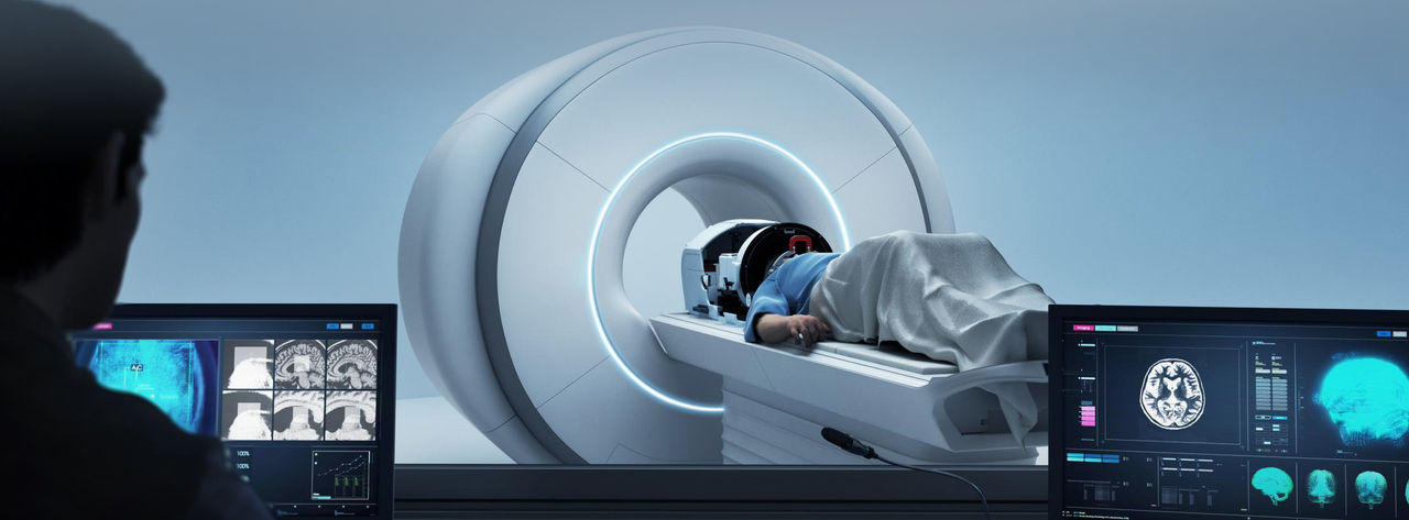
In focused ultrasound, MRI is used to develop a detailed picture of a patient’s brain. Ultrasound wavelengths are then used to heat and kill the specific cells responsible for causing the individual’s tremors, essentially creating a lesion in the region of the brain.
“We can pretreat the region at low levels of heat energy without causing any damage,” Dr. Martello said. “This stuns the cells. We can then review the MRI and ask the patient how they are feeling.”
Adding to the procedure’s safety is the fact that it can be nondestructively tested and guided by immediate patient feedback, according to Dr. Martello. In ET patients, the procedure can be done on both sides of the brain — albeit in separate treatments about nine months apart — to achieve full relief from tremors affecting both sides of the body.
“If the tremor is still there or the patient has side effects, we know we are not in the right spot, but we haven’t done any permanent damage so we can move on to a different area,” Dr. Martello noted. “By doing that, we can make sure we are treating the correct area and avoiding side effects.”
Essential tremor is the most common movement disorder in the United States, affecting an estimated 6.4 million people.1 The disorder typically occurs later in life and is an inherited condition in up to 70% of patients.2
“Everyone has a tremor center of their brain,” Dr. Martello explained. “For patients with tremors it is not functioning correctly.”
Focused ultrasound has been found to be very effective for tremor. A study published in The New England Journal of Medicine in 2016 determined that patients’ mean scores for hand tremor improved by 47% at three months in the cohort receiving the treatment, compared with 0.1% in study participants who underwent a sham procedure.3 And the noninvasive nature of the technique makes it particularly appealing for older patients who may be poorly suited for an invasive approach.
“The results can be dramatic,” Dr. Martello said. “Patients who have been living with this for decades are able to come out of the procedure and drink from a glass.”
Dr. Martello and team will select a very specific area of the brain to create a small lesion to decrease or eliminate the tremor. They have to weigh treating a larger area of the brain for more tremor reduction against possible increased risk of side effects.
Recent five-year data have been promising, showing that Clinical Rating Scale for Tremor scores significantly improved by 73.3% and 73.1% from baseline at both 48 and 60 months post-treatment, respectively.4
“Back in the day, we wanted to treat the whole tremor so we did big lesions, but we saw side effects such as balance issues,” Dr. Martello said, adding that correcting this behavior has caused some surgeons to create too small of a lesion, which may allow for the tremors to return. “Trying to find a middle ground of treating enough of the involved cells, but not so much that you are getting side effects.”
References
- Crawford S, Lally C, Petrillo J, et al. How many adults in the US have essential tremor? Using data from epidemiological studies to derive age-specific estimates of prevalence. Neurology. 2020;94(15 suppl):4458.
- Lorenz D, Frederiksen H, Moises H, et al. High concordance for essential tremor in monozygotic twins of old age. Neurology. 2004;62(2):208-211.
- Elias WJ, Lipsman N, Ondo WG, et al. A randomized trial of focused ultrasound thalamotomy for essential tremor. N Engl J Med. 2016;375(8):730-739.
- Cosgrove GR, Lipsman N, Lozano AM, et al. Magnetic resonance imaging-guided focused ultrasound thalamotomy for essential tremor: 5-year follow-up results. J Neurosurg. 2022;138(4):1028-1033.
A patient diagnosed with multiple sclerosis (MS) 20 years ago would have faced significant disability, pain, and perhaps a shortened life span.
Although there’s currently no cure for the neurologic disorder, which typically occurs in young adults aged 20 to 40 years, neurologists at ChristianaCare are embracing new treatments designed to help manage the course of the disease and limit its disabling effects. Treatments include disease-modifying therapies (DMT), which can slow disease progression and relieve some symptoms.
“The best practice in managing a patient’s MS is through a multidisciplinary team approach that meets both the medical and nonmedical needs of patients, during any stage of the disease,” said Xiaoyang Li, M.D., a neuroimmunologist at ChristianaCare. “Our patients are guided through different treatment options during all stages of their lives, including instruction for patients and families about medicines and how to best manage symptoms.”
Indeed, ChristianaCare’s MS Comprehensive Center of Excellence provides the most effective advanced treatments and symptom management approaches available. Today, MS is considered a chronic disease that is manageable. If it’s diagnosed early and treatment is initiated before symptoms progress, patients can maintain function for years.
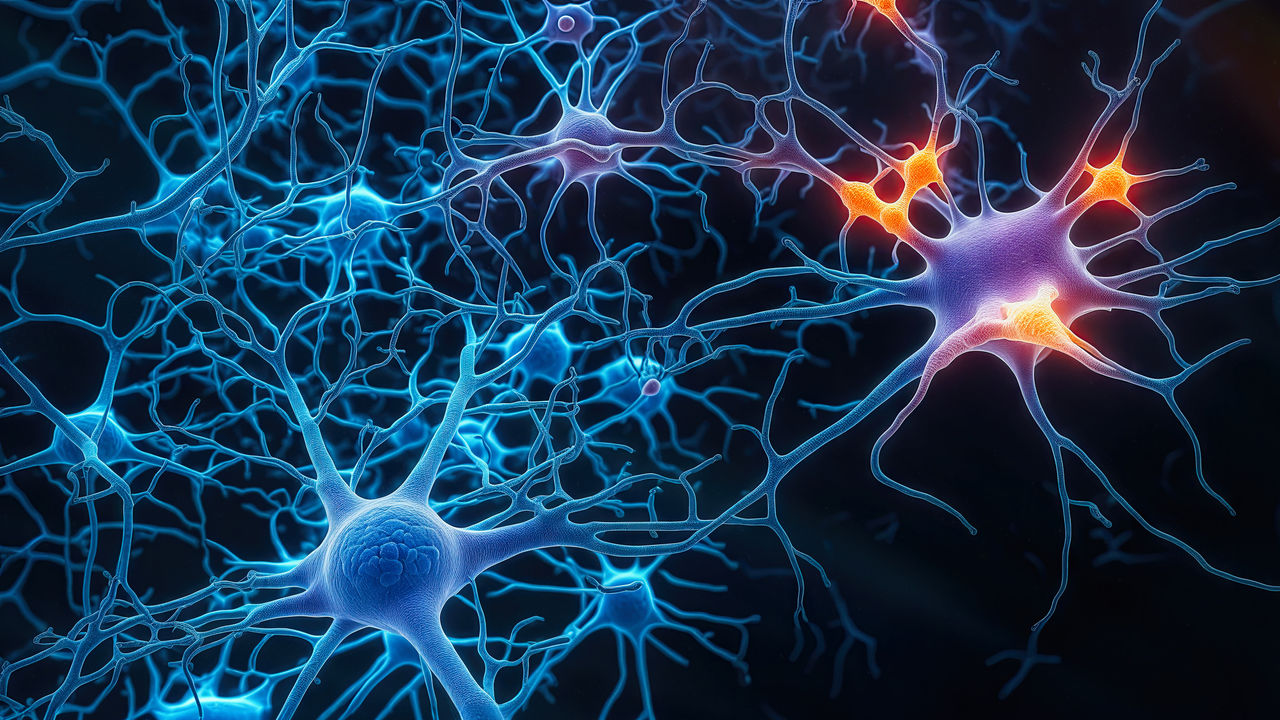
With immunotherapies such as DMTs, patients who start treatment early in the disease course can lead relatively normal lives. In MS, the immune system attacks healthy tissue and nerve cells in error, causing muscle weakness and movement problems. DMTs work by lowering the activity of the immune system, limiting its ability to attack healthy cells. In many patients, these drugs alter the course of MS, reducing their risk for disease relapse, decreasing disease activity and slowing symptom progression.
The FDA has approved more than a dozen DMTs for different types of MS, including clinically isolated syndrome, relapsing-remitting, secondary progressive and primary progressive. However, the drugs have proved most effective in treating the first three types.
Most of the DMTs recommended by neurologists within ChristianaCare’s MS Comprehensive Center of Excellence are infusion-based and can be administered in six-month dosing schedules. Because of a range of choices depending on the patient’s type of MS, treatments can be tailored for a specific patient, allowing for better disease management.
Still, although DMTs offer increased efficacy, often with fewer side effects than more traditional treatments, they do not reverse any nerve damage caused by MS that occurred prior to diagnosis.
Even with these advances, “the most important consideration in the overall management of MS is an early diagnosis and early treatment intervention with the most advanced therapies available,” Dr. Li said.
“On a daily basis, my colleagues and I treat disorders of the blood vessels in the head, neck and spine — aneurysms being one of them,” said Barbara J. Albani, M.D., the medical director for neurointerventional surgery at ChristianaCare. “Given the wide array of devices and techniques that are at our disposal we are able to tailor treatment specifically to each individual patient.”
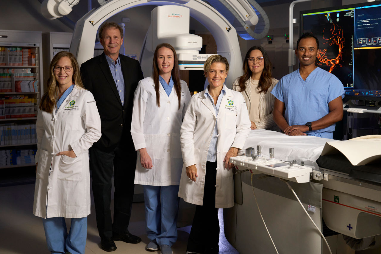
Of these techniques, endovascular coiling is the oldest, dating back to the 1990s. The procedure involves the use of small, flexible catheters that are typically inserted via a patient’s femoral artery. Once the catheter has reached the brain, the surgeon is able to place coils in the aneurysm.
Platelets adhere to the coils and heal in a way that restricts blood from reaching the out-pouching of the aneurysm.1
“This is a very sophisticated way to treat an aneurysm without having to open up the patient’s head and displace their brain,” Dr. Albani said. “Everything is done from within the artery without causing any disruption to the brain tissue.”
Although the technique works on saccular aneurysms — those with a lollipop or mushroom-like shape — others, which are fusiform or wide-necked in shape, require alternative approaches. For these, an open celled stent can be used which has gaps that can be crossed by a wire or a microcatheter, but will hold coils into an aneurysm. The stents are placed in the parent artery near the aneurysm, and a guidewire is passed through the stent in order to place coils within the aneurysm.
“Once placed, the stents help buttress the coils in order to keep them from falling out,” she said.
For certain patients, a finer mesh stent may be used, according to Dr. Albani. These permeable, flow-diverting stents are made of a very fine-gauge mesh that looks similar to a woman’s stocking.
They act to divert blood away from the aneurysm.
“Blood can still seep in, but it is not driving into the aneurysm,” Dr. Albani said. “The analogy I use for patients is it’s kind of like a coffee filter — the water will pass through but not flow with abundance. And the more filters you put in, the less flow you get through it. Over time, the body will endothelialize; the lining of the vessels will grow over the flow-diverting stent, remodeling the blood vessel and effectively sealing it from the inside.”
More recent technologies include the Woven EndoBridge WEB Embolization System (MicroVention) and the Contour Neurovascular System (Stryker).
“The WEB system looks almost like a square ball of yarn and keeps blood from getting into the aneurysm,” Dr. Albani said.
The Contour system, which Dr. Albani noted is shaped like an umbrella that has been inverted by the wind, can be inserted into an aneurysm and cinched down in order to serve as a wall that coils can be inserted behind. Indicated for wide-necked aneurysms, neither system uses a stent, meaning nothing needs to be placed in the parent artery, negating the need for antiplatelet medications that may not work for all patients.
“Whenever we put a stent into somebody’s artery, we have to put them on antiplatelet medications, and this usually involves keeping them on aspirin for life,” Dr. Albani said. “These devices can be a godsend for patients who can’t take aspirin, or who for other reasons we don’t want to put on blood thinners.”
Key to the ChristianaCare team’s approach is a deep understanding of, and experience with, each of the techniques, allowing them to customize each patient’s care.
“We have been early adopters of all of these things, have lots of experience using them and can tailor a patient’s treatment to what they need,” Dr. Albani said. “We have everything we need to make sure the patients are covered, and are here 24/7/365 to help with questions or concerns.”
References
- Ogilvy CS. Neurosurgical clipping versus endovascular coiling of patients with ruptured intracranial aneurysms. Stroke. 2003;34(10):2540-2542.
Patients receiving care in the Neurocritical Care Unit at ChristianaCare with concern for seizure may benefit from a new electroencephalographic (EEG) technology on-boarded by health-system neurologists.
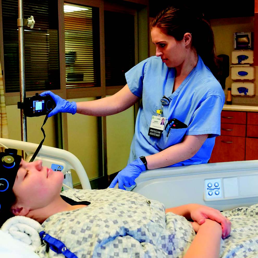
The platform, called Ceribell® EEG (by the manufacturer of the same name), enables rapid evaluation of possible seizure activity when a standard EEG is not immediately. The technology enables ChristianaCare neurologists to quickly evaluate for seizure activity even when EEG technicians are not immediately available.
“This tool allows us to obtain a diagnosis quickly and initiate treatment as needed,” explained Huijun Wang, Ph.D., a specialist in neurology and psychology at ChristianaCare. “It doesn’t have the same sensitivity and specificity as traditional EEG, but it gives us enough information to confirm or rule out seizure in minutes, when we need it, in an emergency situation. That, combined with clinical suspicion, can be enough for us to begin treatment.”
For patients with suspected seizure, time to diagnosis is a key determinant of outcomes, similar to the door-to-needle times in the management of ischemic stroke, as indicated by research. The sooner neurologists can start antiseizure treatment in patients, the more likely they can keep the brain disorder under control.
The primary benefit of Ceribell’s Food and Drug Administration-approved EEG device is that it is completely automated and can be operated by doctors and nurses at the bedside, without a specialized EEG technician, after minimal training. The device is a reduced-montage (8-channel) circumferential EEG that produces tracings covering the frontal, temporal and occipital areas. During evaluation, electrodes are implanted in a soft, adjustable headband attached around the head and secured for electrode grip, and amplified brain signals are transmitted to a pocket-sized device that allows patient data to be entered and viewed in real time.1
Currently, the technology is being used by neurologists as well as nurses and advanced care practitioners in the neuro-ICU at ChristianaCare’s ewark facility. Pending results, it may be expanded to other facilities later this year.
In studies, Ceribell® EEG has been shown to significantly reduce time to diagnosis. A recent analysis published in the journal Epilepsia Open found that mean time to epilepsy diagnosis with the device was 23.8 minutes.2 In her experience with the technology, though, Dr. Wang said it can essentially provide diagnostic information in “real time.”
With that said, Ceribell® EEG doesn’t — and won’t — replace conventional, state-of-the-art EEG platforms, which are more accurate and provide a more complete reading of electrical activity in the brain, according to Richard K. Choi, D.O.. Many patients initially evaluated with the novel device will undergo conventional EEG later to confirm early findings, he said.
“Ceribell® EEG is really like the farmer’s dog barking in the middle of the night — it can warn us that something is or is not happening in a patient’s brain; but the farmer, or in this case the neurologist, will still need to confirm whether it’s something serious,” explained Dr. Choi, a neurologist and neurocritical care specialist at ChristianaCare. “Our use of this technology really highlights ChristianaCare’s ability to see a challenge and find an effective way to address it. It really reflects our values and behaviors, one of which is making sure we’re using all resources available to ensure the best care for our patients.”
References
- Parvizi J, Gururangan K, Razavi B, et al. Detecting silent seizures by their sound. Epilepsia. 2018;59(4):877-884.
- LaMonte MP. Ceribell EEG shortens seizure diagnosis and workforce time and is useful for COVID isolation. Epilepsia Open. 2021;6(2):331-338.
Botox (onabotulinumtoxinA, Allergan) injections have emerged as the most effective option for the treatment of chronic migraine, and specialists at ChristianaCare were early adopters of the innovative approach.
Botox is a neurotoxin that, when injected into muscle tissue, can prevent movement of the treated muscle for a limited time. Best known as a treatment for smoothing wrinkles in the skin on the face, these injections have traditionally been used to treat neuromuscular issues, including spasms and other muscle-contracting conditions.
“In addition to being the most effective treatment we have for refractory chronic migraine, Botox doesn’t have any systemic effects,” said Mitra Assidi-Khansari, M.D., a headache medicine specialist and the director of education in the Division of Neurology. “So, when we inject it into those tiny muscles on the cranial facial area, the medication basically remains in those muscles. It doesn’t float in your system, doesn’t affect the kidney or liver, and doesn’t penetrate your brain and cause cognitive side effects or changes in appetite or mood.”
Migraine headaches are extremely common, particularly in women. Up to 17% of women and 6% of men suffer from migraines.1 Chronic refractory migraine, in which patients experience frequent or long-lasting symptoms for 15 or more days per month, are somewhat less common than episodic migraine, in which patients have symptoms for less than 15 days per month.

As a result, the former “is, unfortunately, a very undertreated disorder,” noted Jessica Bradley, M.D., a headache medicine specialist ChristianaCare. “Many patients don’t seek care until the migraines have gotten bad enough that they are forced to seek treatment options.”
Botox was approved by the FDA in 2010 for use in the treatment of chronic migraine in adults with symptoms that have not responded to three other “standard treatments” (eg, calcitonin gene–related peptide antagonists and receptors). Based on the FDA guidelines, Botox can be administered every three months, for a total of four full treatments annually.
A standard protocol has been established for administering a total of 155 units of Botox. A recommended paradigm calls for injections on the frontal area, the temple, the occipital region over the cervical spinal area and on the top of the shoulders for a total of about 31 shots.
Botox exerts its effect mainly by decreasing signaling on the motor fibers and reducing muscle tension in the cranial facial areas. There are some theories indicating that it can also disrupt the signaling on the sensory fibers.
In a study involving 245 patients with migraine, 82.9% reported that Botox helped control their symptoms.2
Despite its effectiveness, patients need to set expectations on what to anticipate from Botox treatments.
“The first thing I tell patients is that the treatments often don’t work in the first session,” Dr. Bradley said. “Botox treatments often have a cumulative build up in effect, and it can take up to three rounds of treatment to reach maximum migraine control. I explain this to my patients before starting so they know what to expect and won’t feel discouraged if the first treatment does not work well. I encourage patients to try at least three rounds of Botox before determining its effectiveness.”
References
- Stewart WF, Shechter A, Rasmussen BK. Migraine prevalence. A review of population-based studies. Neurology. 1994;44(6 suppl 4):S17-S23.
- Yalinay Dikmen P, Kosak S, Ilgaz Aydinlar E, et al. A single-center retrospective study of onabotulinumtoxinA for treatment of 245 chronic migraine patients: survey results of a real-world experience. Acta Neurol Belg. 2018;118(3):475-484.